What You Should Know:
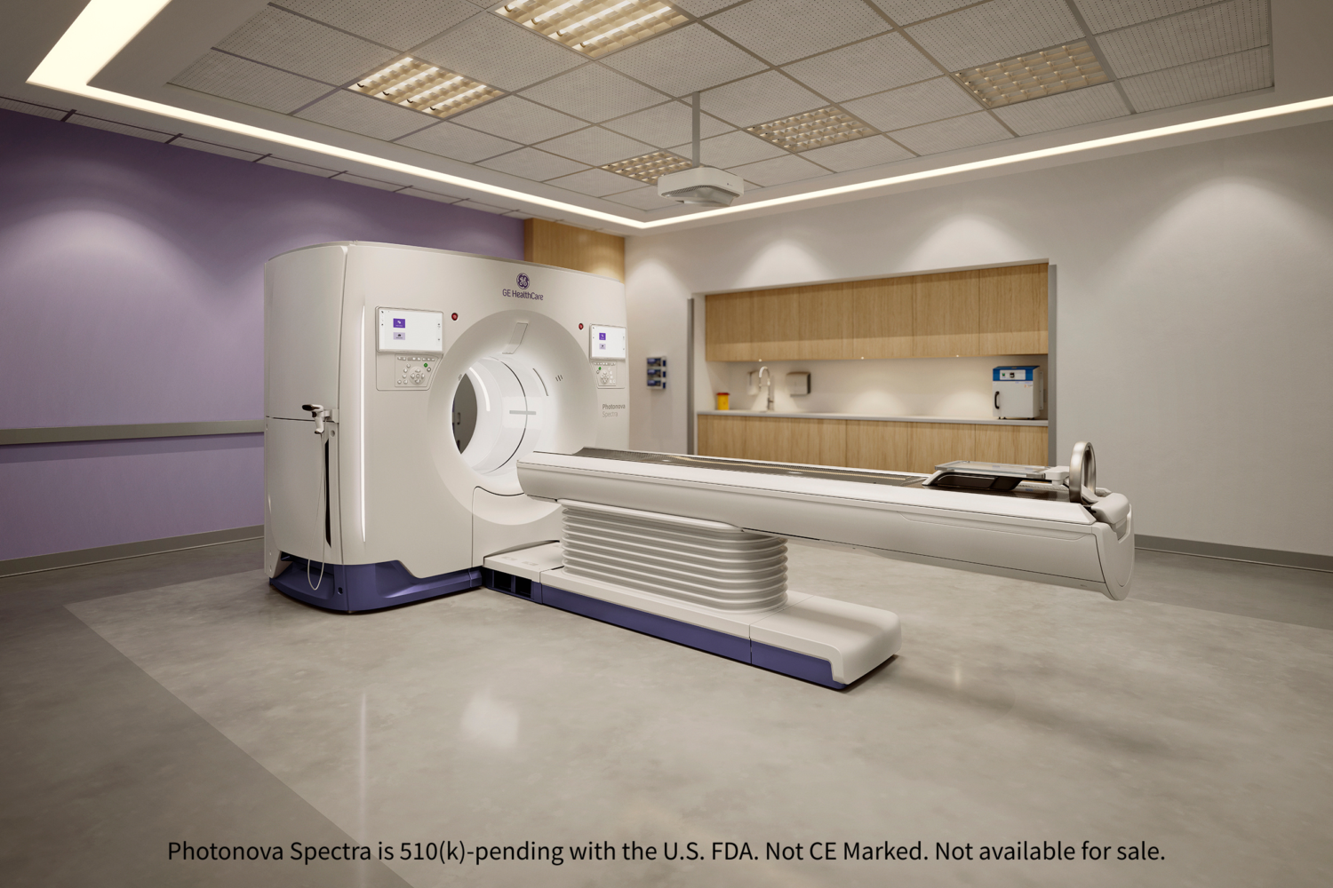
– GE HealthCare has announced the submission of a 510(k) to the U.S. FDA for Photonova Spectra, its new photon-counting computed tomography (PCCT) system with advanced AI algorithms.
– Built on proprietary Deep Silicon detector technology, the system aims to redefine diagnostic confidence by delivering ultra-high-definition (UHD) imaging, enhanced material separation, and rapid acquisition speeds.
Photon Counting CT with Deep Silicon
GE HealthCare’s submission of Photonova Spectra for FDA clearance marks a major milestone in CT innovation. Photon counting CT is a transformative advancement over conventional CT. Unlike older systems that convert X-rays into light, PCCT directly counts individual X-ray photons and measures their energy. This process enables potentially higher spectral and spatial resolution and improved tissue characterization.
Photonova Spectra utilizes Deep Silicon, a proprietary detector material known for its purity and structural consistency. This technology aims to bring enhanced spectral imaging, supporting advanced lesion characterization and treatment monitoring.
Peter Arduini, President & CEO of GE HealthCare, described the system as “more than a new product – it’s a demonstration of what’s possible when vision meets purposeful design,” aiming to “redefine decision-making and care delivery”.
Powering Precision Across Care Pathways
The Photonova Spectra system is intentionally engineered to address rising patient volumes and diagnostic complexity. The system is designed to maximize the vast amounts of data provided—harnessing up to 50 times more data than conventional CT—with the help of NVIDIA’s accelerated computing technology for an effortless workflow with the help of NVIDIA’s accelerated computing technology to enable advanced reconstruction techniques and precise outputs with the aim of supporting enhanced clinical decision-making and smooth workflows.
The clinical potential spans multiple specialties:
- Cardiology: One-second acquisitions support rapid cardiac scans, enabling in-stent lumen assessment and plaque characterization.
- Oncology: Aims for clear lesion characterization and precise quantification, helping clinicians distinguish oncological findings and support treatment monitoring.
- Neurology: Designed for excellent visualization of tiny structures like the inner ear and clear differentiation between brain grey and white matter.